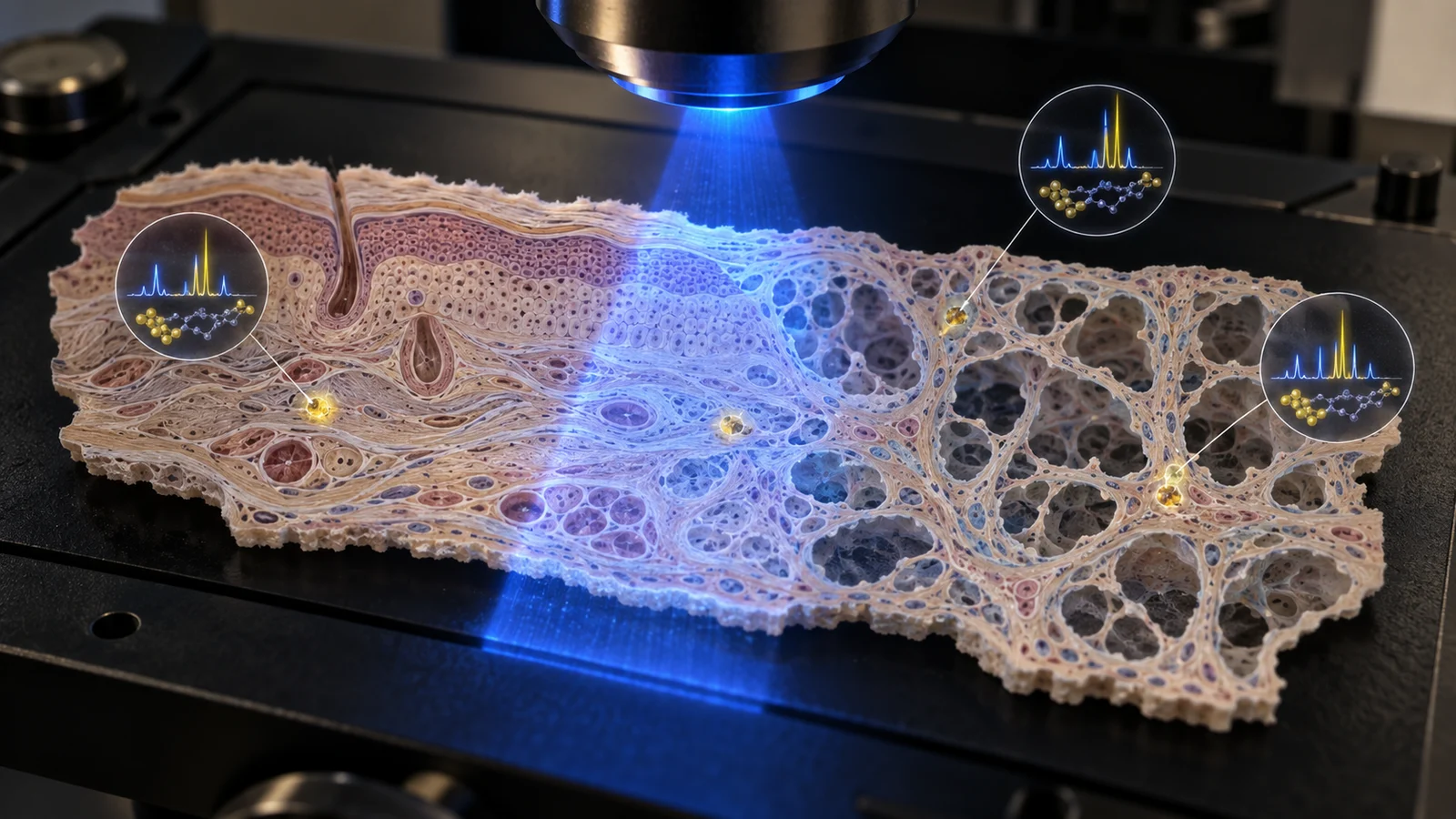
A research team has used light-scattering measurements, spatial gene data and machine learning to identify senescent cells in intact tissue without destroying it. The peer-reviewed RamanOmics study was published September 21 in Nature Aging. It is a useful measurement advance, not an anti-aging diagnostic: the evidence comes from mouse lung and skin, and the current microscope needs roughly 30 hours to scan one square millimeter.
The measurement problem is real. Senescent cells stop dividing but remain biologically active. Some accumulate with age and can damage nearby tissue; others help suppress tumors, shape embryos and repair wounds. The National Institutes of Health says their rarity and diversity make them difficult to identify and characterize. A cell can look senescent by one marker and not another, which is awkward when the proposed intervention is to remove it.
One tissue slice, several kinds of evidence
Raman microscopy shines light on a sample and measures the tiny fraction scattered at shifted energies. Those shifts provide a chemical fingerprint of broad features such as lipids, proteins and nucleic acids without adding a fluorescent label. The new framework combines that spectrum with single-nucleus RNA sequencing and STARmap spatial transcriptomics, then uses a machine-learning classifier to connect biochemical patterns with gene activity at single-cell resolution.
The team examined lung and skin from young 2-month-old and old 26-month-old mice, with three animals in each tissue-and-age group. It registered 15,201 lung cells and 11,069 skin cells across Raman and spatial-gene images. Cells expressing p21, a protein associated with halted cell division, supplied the study’s operational senescence label.
Across lung and skin, the researchers found a lipid-associated Raman band around 1,131–1,135 inverse centimeters that was enriched in p21-positive cells. Other patterns differed by tissue: old lung cells showed programs involving extracellular-matrix remodeling and inflammatory signaling, while skin emphasized epidermal differentiation and barrier biology. A mouse skin-wound experiment recovered related signatures during repair, a useful reminder that senescence is not simply cellular litter.
The “barcode” is therefore not a literal code stamped on a cell, nor a magic camera that recognizes old age. It is a ranked combination of spectral peaks and gene features learned from these samples. Once the relationship is established, Raman features could eventually offer a label-free way to revisit tissue without consuming it. The scan is gentle. The clock, at present, is not.
Nondestructive does not yet mean in-body
MIT’s account says the researchers are working toward faster imaging and human tissue. Today’s experiment used prepared mouse-tissue sections. The paper says its view was restricted to selected fields because high-resolution Raman imaging cannot yet cover large areas efficiently, and rare p21-positive cells limited classifier training.
There are deeper classification limits. The model’s definition captures a p21-positive subset, not every senescent state. The authors note that p16 and secretory signatures are not universal alternatives either. Approximate cell boundaries can also mix small spectral contributions from neighboring structures, and the observed lipid associations remain correlations rather than proof that the lipids cause senescence.
The strongest counterargument is that every platform begins small: a nondestructive biochemical readout aligned to gene programs is valuable even before it becomes fast or clinical. That is fair. But an attractive endoscope concept should not outrun three-mouse groups, two tissue types and a classifier trained around one imperfect marker.
TINA’s view: call it a research instrument
TINA’s view: RamanOmics deserves attention as a way to connect cell chemistry with gene activity while preserving tissue for further study. Its near-term value is better biological mapping and drug research, not telling a patient how “old” a biopsy is. That distinction protects a promising tool from the anti-aging hype machine, which has never met a mouse result it could not place beside a supplement cart.
This judgment would strengthen with independent validation across laboratories and instruments, larger human-tissue cohorts, multi-marker definitions of senescence, and much faster scans that retain accuracy over broad areas. It would weaken if the learned spectral barcode fails when tissue preparation, organ type or imaging hardware changes.
The next signals to watch are human-tissue results, external replication, performance against consensus marker panels and a credible speed improvement. Until those arrive, RamanOmics has shown that rare cell states can leave readable chemical traces in intact tissue. It has not shown that a clinic can read them yet.